| [1] |
Simone L. Ooids: A review[J]. Earth-Science Reviews, 1980, 16: 319-355. |
| [2] |
Richter D K. Calcareous ooids: A synopsis[M]//Peryt T M. Coated grains. Berlin: Springer, 1983: 71-99. |
| [3] |
Peryt T M. Coated grains[M]. Berlin: Springer, 1983: 1-655. |
| [4] |
Beukes N J. Ooids and oolites of the proterophytic Boomplaas Formation, Transvaal Supergroup, Griqualand west, South Africa[M]//Peryt T M. Coated grains. Berlin: Springer, 1983: 199-214. |
| [5] |
Sumner D Y, Grotzinger J P. Numerical modeling of ooid size and the problem of Neoproterozoic giant ooids[J]. Journal of Sedimentary Research, 1993, 63(5): 974-982. |
| [6] |
李飞,王夏,薛武强,等. 一种新的错时相沉积物:巨鲕及其环境意义[J]. 沉积学报,2010,28(3):585-595. |
Li Fei, Wang Xia, Xue Wuqiang, et al. Origin and environmental significance of giant ooids in the Early Triassic: A new kind of anachronistic facies[J]. Acta Sedimentologica Sinica, 2010, 28(3): 585-595. |
| [7] |
Tucker M E, Wright V P. Carbonate sedimentology[M]. Oxford: Blackwell Science, 1990: 1-496. |
| [8] |
Siahi M, Hofmann A, Master S, et al. Carbonate ooids of the Mesoarchaean Pongola Supergroup, South Africa[J]. Geobiology, 2017, 15(6): 750-766. |
| [9] |
Hofmann H J, Grey K, Hickman A H, et al. Origin of 3.45 Ga coniform stromatolites in Warrawoona Group, western Australia[J]. GSA Bulletin, 1999, 111(8): 1256-1262. |
| [10] |
Wilkinson B H, Owen R M, Carroll A R. Submarine hydrothermal weathering, global eustasy, and carbonate polymorphism in Phanerozoic marine oolites[J]. Journal of Sedimentary Research, 1985, 55(2): 171-183. |
| [11] |
Li F, Yan J X, Chen Z Q, et al. Global oolite deposits across the Permian-Triassic boundary: A synthesis and implications for palaeoceanography immediately after the end-Permian biocrisis[J]. Earth-Science Reviews, 2015, 149: 163-180. |
| [12] |
Opdyke B N, Wilkinson B H. Paleolatitude distribution of Phanerozoic marine ooids and cements[J]. Palaeogeography, Palaeoclimatology, Palaeoecology, 1990, 78(1/2): 135-148. |
| [13] |
Lees A, Buller A T. Modern temperate-water and warm-water shelf carbonate sediments contrasted[J]. Marine Geology, 1972, 13(5): M67-M73. |
| [14] |
Lippmann F. Sedimentary carbonate minerals[M]. Berlin: Springer, 1973: 1-229. |
| [15] |
Gallagher S J, Reuning L, Himmler T, et al. The enigma of rare Quaternary oolites in the Indian and Pacific Oceans: A result of global oceanographic physicochemical conditions or a sampling bias?[J]. Quaternary Science Reviews, 2018, 200: 114-122. |
| [16] |
Reeder S L, Rankey E C. Interactions between tidal flows and ooid shoals, northern Bahamas[J]. Journal of Sedimentary Research, 2008, 78(3): 175-186. |
| [17] |
Ward W C, Brady M J. High-energy carbonates on the inner shelf, northeastern Yucatan Peninsula, Mexico[J]. GCAGS Transactions, 1973, 23: 226-238. |
| [18] |
Trower E J, Cantine M D, Gomes M L, et al. Active ooid growth driven by sediment transport in a high-energy shoal, Little Ambergris Cay, Turks and Caicos Islands[J]. Journal of Sedimentary Research, 2018, 88(9): 1132-1151. |
| [19] |
Purser B H. The Persian Gulf: Holocene carbonate sedimentation and diagenesis in a shallow Epicontinental sea[M]. Berlin: Springer, 1973: 1-474. |
| [20] |
Logan B W, Davies G R, Read J F, et al. Carbonate sedimentation and environments, Shark Bay, western Australia[M]. Tulsa: American Association of Petroleum Geologists, 1970: 1-205. |
| [21] |
Rankey E C, Reeder S L. Holocene ooids of Aitutaki Atoll, Cook Islands, South Pacific[J]. Geology, 2009, 37(11): 971-974. |
| [22] |
Witzke B J. Palaeoclimatic constraints for Palaeozoic palaeolatitudes of Laurentia and Euramerica[M]//McKerrow W S, Scotese C R. Palaeozoic palaeogeography and biogeography. London: Geological Society, 1990: 57-73. |
| [23] |
Li F, Gong Q L, Burne R V, et al. Ooid factories operating under hothouse conditions in the earliest Triassic of South China[J]. Global and Planetary Change, 2019, 172: 336-354. |
| [24] |
Duguid S M A, Kyser T K, James N P, et al. Microbes and ooids[J]. Journal of Sedimentary Research, 2010, 80(3): 236-251. |
| [25] |
Beaupré S R, Roberts M L, Burton J R, et al. Rapid, high-resolution 14C chronology of ooids[J]. Geochimica et Cosmochimica Acta, 2015, 159: 126-138. |
| [26] |
Bathurst R G C. Carbonate sediments and their diagenesis[M]. Amsterdam: Elsevier Science, 1972: 1-658. |
| [27] |
李飞,武思琴,刘柯. 鲕粒原生矿物识别及对海水化学成分变化的指示意义[J]. 沉积学报,2015,33(3):500-511. |
Li Fei, Wu Siqin, Liu Ke. Identification of ooid primary mineralogy: A clue for understanding the variation in paleo-oceanic chemistry[J]. Acta Sedimentologica Sinica, 2015, 33(3): 500-511. |
| [28] |
宋文天,刘建波. 碳酸盐鲕粒包壳结构研究综述[J]. 古地理学报,2020,22(1):147-160. |
Song Wentian, Liu Jianbo. A review of cortical structures of carbonate ooids[J]. Journal of Palaeogeography, 2020, 22(1): 147-160. |
| [29] |
Sandberg P A. An oscillating trend in Phanerozoic non-skeletal carbonate mineralogy[J]. Nature, 1983, 305(5929): 19-22. |
| [30] |
Hooke R. Micrographia, or, Some physiological descriptions of minute bodies made by magnifying glasses, with observations and inquiries thereupon[M]. London: J. Martyn and J. Allestry, 1665: 1-246. |
| [31] |
Brückmann F E. Specimen physicum exhibens historiam naturalem, oolithi seu ovariorum piscium & concharum in saxa mutatorum[M]. Helmestadii: Salomoni & Schnorrii, 1721: 1-28. |
| [32] |
Da Costa E M. A natural history of fossils[M]. London: L. Davis and C. Reymers, 1757: 1-294. |
| [33] |
De Saussure H B. Voyages dans les Alpes, précédés d'un essai sur l'histoire naturelle des environs de Genève[M]. Neuchâtel: Franche-Borel, 1779: 1-540. |
| [34] |
Burne R V, Eade J C, Paul J. The natural history of ooliths: Franz Ernst Brückmann’s treatise of 1721 and its significance for the understanding of oolites[J]. Hallesches Jahrbuch für Geowissenschaften, 2012, 34: 93-114. |
| [35] |
Sorby H C. The structure and origin of limestones[J]. Proceedings of the Geological Society of London, 1879, 35: 56-95. |
| [36] |
Wethered E. On the occurrence of the genus Girvanella in oolitic rocks, and remarks in oolitic structure[J]. Quarterly Journal of the Geological Society, 1890, 46(1/2/3/4): 270-283. |
| [37] |
Rothpletz A. On the formation of oolite[J]. Botanisches Centralblatt, 1892, 10(5): 279-283. |
| [38] |
Linck G. Die bildung der oolithe und rogensteine[J]. Neues Jahrbuch für Geologie und Paläontologie Abhandlungen, 1903, 16: 495-513. |
| [39] |
Drew G H. The action of some denitrifying bacteria in tropical and temperate seas, and the bacterial precipitation of calcium carbonate in the sea[J]. Journal of the Marine Biological Association of the United Kingdom, 1911, 9(2): 142-155. |
| [40] |
Vaughan T W. Preliminary remarks on the geology of the Bahamas, with special reference to the origin of the Bahaman and Floridian oolites[J]. Washington: Tortugas Laboratory of the Carnegie Institution of Washington, 1914, 102: 47-54. |
| [41] |
Monaghan P H, Lytle M L. The origin of calcareous ooliths[J]. Journal of Sedimentary Research, 1956, 26(2): 111-118. |
| [42] |
Newell N D, Purdy E G, Imbrie J. Bahamian oölitic sand[J]. The Journal of Geology, 1960, 68(5): 481-497. |
| [43] |
Fabricius F H. Origin of marine oöids and grapestones[M]. Stuttgart: E. Schweizerbart'sche Verlagsbuchhandlung, 1977: 1-113. |
| [44] |
Gerdes G, Dunajtschik-Piewak K, Riege H, et al. Structural diversity of biogenic carbonate particles in microbial mats[J]. Sedimentology, 1994, 41(6): 1273-1294. |
| [45] |
Reitner J, Arp G, Thiel V, et al. Organic matter in Great Salt Lake ooids (Utah, USA) - First approach to a formation via organic matrices[J]. Facies, 1997, 36: 210-219. |
| [46] |
Folk R L. SEM imaging of bacteria and nannobacteria in carbonate sediments and rocks[J]. Journal of Sedimentary Research, 1993, 63(5): 990-999. |
| [47] |
Diaz M R, Eberli G P, Blackwelder P, et al. Microbially mediated organomineralization in the formation of ooids[J]. Geology, 2017, 45(9): 771-774. |
| [48] |
Pacton M, Ariztegui D, Wacey D, et al. Going nano: A new step toward understanding the processes governing freshwater ooid formation[J]. Geology, 2012, 40(6): 547-550. |
| [49] |
Plee K, Ariztegui D, Martini R, et al. Unravelling the microbial role in ooid formation - results of an in situ experiment in modern freshwater Lake Geneva in Switzerland[J]. Geobiology, 2008, 6(4): 341-350. |
| [50] |
Plée K, Pacton M, Ariztegui D. Discriminating the role of photosynthetic and heterotrophic microbes triggering low-Mg calcite precipitation in freshwater biofilms (Lake Geneva, Switzerland)[J]. Geomicrobiology Journal, 2010, 27(5): 391-399. |
| [51] |
Summons R E, Bird L R, Gillespie A L, et al. Lipid biomarkers in ooids from different locations and ages: Evidence for a common bacterial flora[J]. Geobiology, 2013, 11(5): 420-436. |
| [52] |
Diaz M R, Van Norstrand J D, Eberli G P, et al. Functional gene diversity of oolitic sands from Great Bahama Bank[J]. Geobiology, 2014, 12(3): 231-249. |
| [53] |
Diaz M R, Eberli G P. Decoding the mechanism of formation in marine ooids: A review[J]. Earth-Science Reviews, 2019, 190: 536-556. |
| [54] |
Budd D A, Land L S. Geochemical imprint of meteoric diagenesis in Holocene ooid sands, Schooner Cays, Bahamas: Correlation of calcite cement geochemistry with extant groundwaters[J]. Journal of Sedimentary Research, 1990, 60(3): 361-378. |
| [55] |
Heydari E, Snelling R D, Dawson W C, et al. Ooid mineralogy and diagenesis of the Pitkin Formation, North-central Arkansas[M]//Keith B D, Zuppann C W. Mississippian oolites and modern analogs. Tulsa: American Association of Petroleum Geologists, 1993: 175-184. |
| [56] |
Kidder D L, Hall S. Petrology and diagenetic evolution of Neoproterozoic ooids (Libby Formation, western Montana, U.S.A.)[J]. Precambrian Research, 1993, 63(1/2): 83-96. |
| [57] |
Harris P M. Facies anatomy and diagenesis of a Bahamian ooid shoal[M]. Miami: University of Miami, 1979: 1-150. |
| [58] |
Li F, Webb G E, Algeo T J, et al. Modern carbonate ooids preserve ambient aqueous REE signatures[J]. Chemical Geology, 2019, 509: 163-177. |
| [59] |
Kalkowsky E. Oolith und Stromatolith im norddeutschen Buntsandstein[J]. Zeitschrift der Deutschen Geologischen Gesellschaft Band, 1908, 60: 68-125. |
| [60] |
Batchelor M T, Burne R V, Henry B I, et al. A biofilm and organomineralisation model for the growth and limiting size of ooids[J]. Scientific Reports, 2018, 8(1): 559. |
| [61] |
Dupraz C, Reid R P, Braissant O, et al. Processes of carbonate precipitation in modern microbial mats[J]. Earth-Science Reviews, 2009, 96(3): 141-162. |
| [62] |
Brown T C. Origin of oolites and the oolitic texture in rocks[J]. GSA Bulletin, 1914, 25(1): 745-780. |
| [63] |
Illing L V. Bahaman calcareous sands[J]. AAPG Bulletin, 1954, 38(1): 1-95. |
| [64] |
Eardley A J. Sediments of Great Salt Lake, Utah[J]. AAPG Bulletin, 1938, 22(10): 1305-1411. |
| [65] |
Harris P, Diaz M R, Eberli G P. The formation and distribution of modern ooids on Great Bahama Bank[J]. Annual Review of Marine Science, 2019, 11: 491-516. |
| [66] |
梅冥相. 鲕粒成因研究的新进展[J]. 沉积学报,2012,30(1):20-32. |
Mei Mingxiang. Brief introduction on new advances on the origin of ooids[J]. Acta Sedimentologica Sinica, 2012, 30(1): 20-32. |
| [67] |
Bandy M C, Bandy J A. De natura fossilium (textbook of mineralogy)[M]. New York: The Geological Society, 1955: 1-240. |
| [68] |
De La Beche H T. The geological observer[M]. Philadelphia: Blanchard and Lea, 1851: 1-695. |
| [69] |
Dana J D. Corals and coral islands[M]. New York: Dodd Mead & Co, 1890: 1-398. |
| [70] |
Bathurst R G C. Precipitation of oöids and other aragonite fabrics in warm seas[M]//Müller G, Friedman G M. Recent developments in carbonate sedimentology in Central Europe. Berlin: Springer, 1968: 1-10. |
| [71] |
Land L S, Behrens E W, Frishman S A. The ooids of Baffin Bay, Texas[J]. Journal of Sedimentary Research, 1979, 49(4): 1269-1277. |
| [72] |
Morse J W, Mackenzie F T. Geochemistry of sedimentary carbonates[M]. Amsterdam: Elsevier, 1990: 1-725. |
| [73] |
Shearman D J, Twyman J, Karimi M Z. The genesis and diagenesis of oolites[J]. Proceedings of the Geologists' Association, 1970, 81(3): 561-564, IN7-IN9, 565-575. |
| [74] |
Folk R L. Carbonate petrography in the post-Sorbian age[M]//Ginsburg R N. Evolving concepts in sedimentology. Baltimore: Johns Hopkins University Press, 1973: 118-158. |
| [75] |
Wethered E B. The formation of oolite[J]. Quarterly Journal of the Geological Society, 1895, 51(1/2/3/4): 196-209. |
| [76] |
Spincer B R. Oolitized fragments of filamentous calcimicrobes and the pseudofossil affinity of Nuia Maslov from the Upper Cambrian rocks of central Texas[J]. Journal of Paleontology, 1998, 72(3): 577-584. |
| [77] |
Liu W, Zhang X L. Girvanella-coated grains from Cambrian oolitic limestone[J]. Facies, 2012, 58(4): 779-787. |
| [78] |
代明月,齐永安,陈尧,等. 豫西渑池地区寒武系第三统张夏组的巨鲕及其成因[J]. 古地理学报,2014,16(5):726-734. |
Dai Mingyue, Qi Yong’an, Chen Yao, et al. Giant ooids and their genetic analysis from the Zhangxia Formation of Cambrian Series 3 in Mianchi area, western Henan province[J]. Journal of Palaeogeography, 2014, 16(5): 726-734. |
| [79] |
Han Z Z, Zhang X L, Chi N J, et al. Cambrian oncoids and other microbial-related grains on the North China Platform[J]. Carbonates and Evaporites, 2015, 30(4): 373-386. |
| [80] |
Li F, Yan J X, Burne R V, et al. Paleo-seawater REE compositions and microbial signatures preserved in laminae of Lower Triassic ooids[J]. Palaeogeography, Palaeoclimatology, Palaeoecology, 2017, 486: 96-107. |
| [81] |
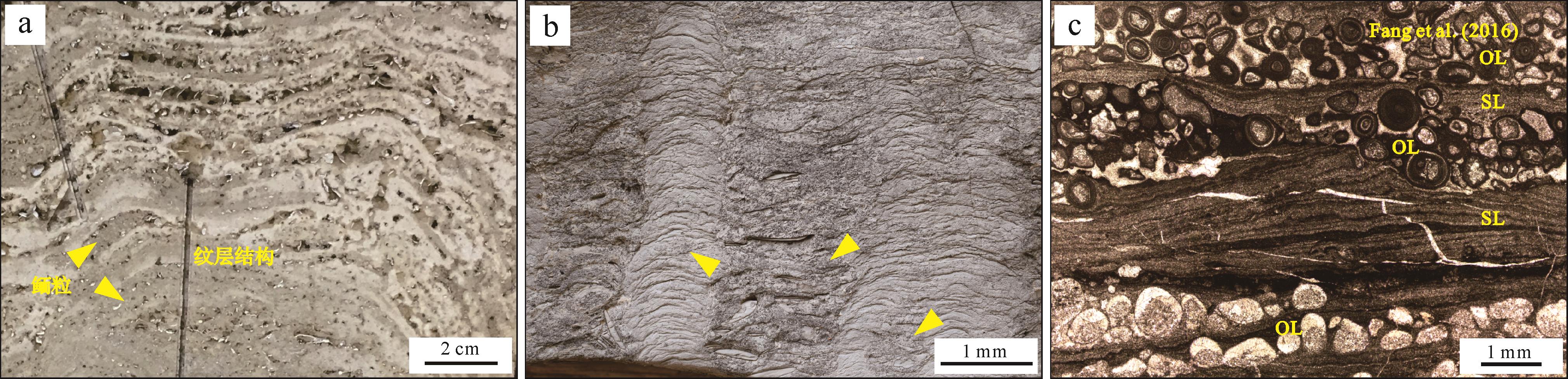
Fang Y H, Chen Z Q, Kershaw S, et al. An Early Triassic (Smithian) stromatolite associated with giant ooid banks from Lichuan (Hubei province), South China: Environment and controls on its formation[J]. Palaeogeography, Palaeoclimatology, Palaeoecology, 2017, 486: 108-122. |
| [82] |
Cayeux L. Les roches sédimentaires de France: Roches carbonatées[M]. Paris: Masson, 1935: 1-463. |
| [83] |
Sandberg P A. New interpretations of Great Salt Lake ooids and of ancient non-skeletal carbonate mineralogy[J]. Sedimentology, 1975, 22(4): 497-537. |
| [84] |
Klement K W, Toomey D F. Role of the blue-green alga Girvanella in skeletal grain destruction and lime-mud formation in the Lower Ordovician of West Texas[J]. Journal of Sedimentary Research, 1967, 37(4): 1045-1051. |
| [85] |
Golubic S, Seong-Joo L, Browne K M. Cyanobacteria: Architects of sedimentary structures[M]//Riding R E, Awramik S M. Microbial sediments. Berlin: Springer, 2000: 57-67. |
| [86] |
Shearman D J, P A d'E Skipwith. Organic matter in recent and ancient limestones and its role in their diagenesis[J]. Nature, 1965, 208(5017): 1310-1311. |
| [87] |
Mitterer R M. Amino acid composition of organic matrix in calcareous oolites[J]. Science, 1968, 162(3861): 1498-1499. |
| [88] |
Davies P J, Bubela B, Ferguson J. The formation of ooids[J]. Sedimentology, 1978, 25(5): 703-730. |
| [89] |
Ferguson J, Bubela B, Davies P J. Synthesis and possible mechanism of formation of radial carbonate ooids[J]. Chemical Geology, 1978, 22: 285-308. |
| [90] |
Reid R P, Macintyre I G. Microboring versus recrystallization: Further insight into the micritization process[J]. Journal of Sedimentary Research, 2000, 70(1): 24-28. |
| [91] |
Drew G H. On the precipitation of calcium carbonate in the sea by marine bacteria, and on the action of denitrifying bacteria in tropical and temperate seas[J]. Journal of the Marine Biological Association of the United Kingdom, 1913, 9(4): 479-524. |
| [92] |
Mitterer R M. Biogeochemistry of aragonite mad and oolites[J]. Geochimica et Cosmochimica Acta, 1972, 36(12): 1407-1422. |
| [93] |
Suess E, Futterer D. Aragonitic ooids: Experimental precipitation from seawater in the presence of humic acid[J]. Sedimentology, 1972, 19(1/2): 129-139. |
| [94] |
Trichet J. Etude de la composition de la fraction organique des oolites. Comparaison avec celle des membranes des bactéries et des cyanophycées[J]. Comptes rendus de l'Académie des Sciences de Paris, Série D, 1968, 267: 1492-1494. |
| [95] |
Krumbein W E. Photolithotropic and chemoorganotrophic activity of bacteria and algae as related to beachrock formation and degradation (gulf of Aqaba, Sinai)[J]. Geomicrobiology Journal, 1979, 1(2): 139-203. |
| [96] |
Krumbein W E. Calcification by bacteria and algae[M]//Trudinger P A, Swaine D J. Biogeochemical cycling of mineral-forming elements. Amsterdam: Elsevier, 1979: 47-68. |
| [97] |
Krumbein W E, Cohen Y, Shilo M. Solar Lake (Sinai). 4. Stromatolitic cyanobacterial mats[J]. Limnology and Oceanography, 1977, 22(4): 635-656. |
| [98] |
Decho A W. Microbial exopolymer secretions in ocean environments: Their role(s) in food webs and marine processes[J]. Oceanography and Marine Biology: An Annual Review, 1990, 28: 73-153. |
| [99] |
Decho A W, Visscher P T, Reid R P. Production and cycling of natural microbial exopolymers (EPS) within a marine stromatolite[J]. Palaeogeography, Palaeoclimatology, Palaeoecology, 2005, 219(1/2): 71-86. |
| [100] |
Gallagher K L, Dupraz C, Braissant O, et al. Mineralization of sedimentary biofilms: Modern mechanistic insights[M]//Bailey W C. Biofilms: Formation, development and properties. New York: Nova Science Publishers, 2010: 227-258. |
| [101] |
Folk R L, Leo Lynch F. Organic matter, putative nannobacteria and the formation of ooids and hardgrounds[J]. Sedimentology, 2001, 48(2): 215-229. |
| [102] |
Folk R L, Chafetz H S. Bacterially induced microscale and nanoscale carbonate precipitates[M]//Riding R E, Awramik S M. Microbial sediments. Berlin: Springer, 2000: 40-49. |
| [103] |
Young J D, Martel J. The rise and fall of nanobacteria[J]. Scientific American, 2010, 302(1): 52-59. |
| [104] |
Southam G. A structural comparison of bacterial microfossils vs. 'nanobacteria' and nanofossils[J]. Earth-Science Reviews, 1999, 48(4): 251-264. |
| [105] |
Obst M, Dynes J J, Lawrence J R, et al. Precipitation of amorphous CaCO3 (aragonite-like) by cyanobacteria: A STXM study of the influence of EPS on the nucleation process[J]. Geochimica et Cosmochimica Acta, 2009, 73(14): 4180-4198. |
| [106] |
Benzerara K, Menguy N, López-García P, et al. Nanoscale detection of organic signatures in carbonate microbialites[J]. Proceedings of the National Academy of Sciences of the United States of America, 2006, 103(25): 9440-9445. |
| [107] |
Couradeau E, Benzerara K, Gérard E, et al. An early-branching microbialite cyanobacterium forms intracellular carbonates[J]. Science, 2012, 336(6080): 459-462. |
| [108] |
Diaz M R, Swart P K, Eberli G P, et al. Geochemical evidence of microbial activity within ooids[J]. Sedimentology, 2015, 62(7): 2090-2112. |
| [109] |
Konhauser K, Riding R. Bacterial biomineralization[M]//Knoll A H, Canfield D E, Konhauser K O. Fundamentals of geobiology. Chichester: John Wiley & Sons, 2012: 105-130. |
| [110] |
Castanier S, Le Métayer-Levrel G, Perthuisot J P. Bacterial roles in the precipitation of carbonate minerals[M]//Riding R E, Awramik S M. Microbial sediments. Berlin: Springer, 2000: 32-39. |
| [111] |
Visscher P T, Stolz J F. Microbial mats as bioreactors: Populations, processes, and products[J]. Palaeogeography, Palaeoclimatology, Palaeoecology, 2005, 219(1/2): 87-100. |
| [112] |
Riding R. Calcified cyanobacteria[M]//Reitner J, Thiel V. Encyclopedia of geobiology. Dordrecht: Springer, 2011: 211-223. |
| [113] |
Riding R. Temporal variation in calcification in marine cyanobacteria[J]. Journal of the Geological Society, 1992, 149(6): 979-989. |
| [114] |
Davaud E, Girardclos S. Recent freshwater ooids and oncoids from western Lake Geneva (Switzerland): Indications of a common organically mediated origin[J]. Journal of Sedimentary Research, 2001, 71(3): 423-429. |
| [115] |
Arp G, Reimer A, Reitner J. Photosynthesis-induced biofilm calcification and calcium concentrations in Phanerozoic oceans[J]. Science, 2001, 292(5522): 1701-1704. |
| [116] |
Dupraz C, Visscher P T. Microbial lithification in marine stromatolites and hypersaline mats[J]. Trends in Microbiology, 2005, 13(9): 429-438. |
| [117] |
Arp G, Reimer A, Reitner J. Microbialite formation in seawater of increased alkalinity, Satonda Crater Lake, Indonesia[J]. Journal of Sedimentary Research, 2003, 73(1): 105-127. |
| [118] |
Couradeau E, Benzerara K, Gérard E, et al. Cyanobacterial calcification in modern microbialites at the submicrometer scale[J]. Biogeosciences, 2013, 10(8): 5255-5266. |
| [119] |
Wacey D, Urosevic L, Saunders M, et al. Mineralisation of filamentous cyanobacteria in Lake Thetis stromatolites, western Australia[J]. Geobiology, 2018, 16(2): 203-215. |
| [120] |
Burne R V, Moore L S, Christy A G, et al. Stevensite in the modern thrombolites of Lake Clifton, western Australia: A missing link in microbialite mineralization?[J]. Geology, 2014, 42(7): 575-578. |
| [121] |
Souza-Egipsy V, Wierzchos J, Ascaso C, et al. Mg–silica precipitation in fossilization mechanisms of sand tufa endolithic microbial community, Mono Lake (California)[J]. Chemical Geology, 2005, 217(1/2): 77-87. |
| [122] |
Pace A, Bourillot R, Bouton A, et al. Formation of stromatolite lamina at the interface of oxygenic-anoxygenic photosynthesis[J]. Geobiology, 2018, 16(4): 378-398. |
| [123] |
Riding R. Cyanobacterial calcification, carbon dioxide concentrating mechanisms, and Proterozoic–Cambrian changes in atmospheric composition[J]. Geobiology, 2006, 4(4): 299-316. |
| [124] |
Riding R. An atmospheric stimulus for cyanobacterial-bioinduced calcification ca. 350 million years ago?[J]. PALAIOS, 2009, 24(10): 685-696. |
| [125] |
Huisman J, Codd G A, Paerl H W, et al. Cyanobacterial blooms[J]. Nature Reviews Microbiology, 2018, 16(8): 471-483. |
| [126] |
Raven J A, Beardall J, Sánchez-Baracaldo P. The possible evolution and future of CO2-concentrating mechanisms[J]. Journal of Experimental Botany, 2017, 68(14): 3701-3716. |
| [127] |
Tang D J, Shi X Y, Shi Q, et al. Organomineralization in Mesoproterozoic giant ooids[J]. Journal of Asian Earth Sciences, 2015, 107: 195-211. |
| [128] |
Soetaert K, Hofmann A F, Middelburg J J, et al. Reprint of “The effect of biogeochemical processes on pH”[J]. Marine Chemistry, 2007, 106(1/2): 380-401. |
| [129] |
Canfield D E, Thamdrup B. Towards a consistent classification scheme for geochemical environments, or, why we wish the term 'suboxic' would go away[J]. Geobiology, 2009, 7(4): 385-392. |
| [130] |
Dupraz, C, Visscher, P T, Baumgartner, L K, Reid, R P. Microbe–mineral interactions: early carbonate precipitation in a hypersaline lake (Eleuthera Island, Bahamas)[J]. Sedimentology, 2004, 51(4): 745-765. |
| [131] |
Diaz M R, Piggot A M, Eberli G P, et al. Bacterial community of oolitic carbonate sediments of the Bahamas Archipelago[J]. Marine Ecology Progress Series, 2013, 485: 9-24. |
| [132] |
Erşan Y Ç, De Belie N, Boon N. Microbially induced CaCO3 precipitation through denitrification: An optimization study in minimal nutrient environment[J]. Biochemical Engineering Journal, 2015, 101: 108-118. |
| [133] |
Hamdan N, Kavazanjian Jr E, Rittmann B E, et al. Carbonate mineral precipitation for soil improvement through microbial denitrification[J]. Geomicrobiology Journal, 2017, 34(2): 139-146. |
| [134] |
Rassmann J, Lansard B, Pozzato L, et al. Carbonate chemistry in sediment porewaters of the Rhône River delta driven by early diagenesis (northwestern Mediterranean)[J]. Biogeosciences, 2016, 13(18): 5379-5394. |
| [135] |
Canfield D E, Des Marais D J. Biogeochemical cycles of carbon, sulfur, and free oxygen in a microbial mat[J]. Geochimica et Cosmochimica Acta, 1993, 57(16): 3971-3984. |
| [136] |
Lyons W B, Long D T, Hines M E, et al. Calcification of cyanobacterial mats in Solar Lake, Sinai[J]. Geology, 1984, 12(10): 623-626. |
| [137] |
Castanier S, Le Métayer-Levrel G, Perthuisot J P. Ca-carbonates precipitation and limestone genesis — the microbiogeologist point of view[J]. Sedimentary Geology, 1999, 126(1/2/3/4): 9-23. |
| [138] |
Braissant O, Decho A W, Dupraz C, et al. Exopolymeric substances of sulfate-reducing bacteria: Interactions with calcium at alkaline pH and implication for formation of carbonate minerals[J]. Geobiology, 2007, 5(4): 401-411. |
| [139] |
Wright D T. The role of sulphate-reducing bacteria and cyanobacteria in dolomite formation in distal ephemeral lakes of the Coorong region, South Australia[J]. Sedimentary Geology, 1999, 126(1/2/3/4): 147-157. |
| [140] |
Canfield D E, Des Marais D J. Aerobic sulfate reduction in microbial mats[J]. Science, 1991, 251(5000): 1471-1473. |
| [141] |
Fründ C, Cohen Y. Diurnal cycles of sulfate reduction under oxic conditions in cyanobacterial mats[J]. Applied and Environmental Microbiology, 1992, 58(1): 70-77. |
| [142] |
Baumgartner L K, Reid R P, Dupraz C, et al. Sulfate reducing bacteria in microbial mats: Changing paradigms, new discoveries[J]. Sedimentary Geology, 2006, 185(3/4): 131-145. |
| [143] |
Teske A, Ramsing N B, Habicht K, et al. Sulfate-reducing bacteria and their activities in cyanobacterial mats of Solar Lake (Sinai, Egypt)[J]. Applied and Environmental Microbiology, 1998, 64(8): 2943-2951. |
| [144] |
Caumette P, Matheron R, Raymond N, et al. Microbial mats in the hypersaline ponds of Mediterranean salterns (Salins-de-Giraud, France)[J]. FEMS Microbiology Ecology, 1994, 13(4): 273-286. |
| [145] |
O'reilly S S, Mariotti G, Winter A R, et al. Molecular biosignatures reveal common benthic microbial sources of organic matter in ooids and grapestones from Pigeon Cay, The Bahamas[J]. Geobiology, 2017, 15(1): 112-130. |
| [146] |
Revsbech N P, Jorgensen B B, Blackburn T H, et al. Microelectrode studies of the photosynthesis and O2, H2S, and pH profiles of a microbial mat[J]. Limnology and Oceanography, 1983, 28(6): 1062-1074. |
| [147] |
Visscher P T, Prins R A, Van Gemerden H. Rates of sulfate reduction and thiosulfate consumption in a marine microbial mat[J]. FEMS Microbiology Letters, 1992, 86(4): 283-293. |
| [148] |
Edgcomb V P, Bernhard J M, Beaudoin D, et al. Molecular indicators of microbial diversity in oolitic sands of Highborne Cay, Bahamas[J]. Geobiology, 2013, 11(3): 234-251. |
| [149] |
Anderson N T, Cowan C A, Bergmann K D. A case for the growth of ancient ooids within the sediment pile[J]. Journal of Sedimentary Research, 2020, 90: 843-854. |
| [150] |
Mariotti G, Pruss S B, Summons R E, et al. Contribution of benthic processes to the growth of ooids on a low-energy shore in Cat Island, The Bahamas[J]. Minerals, 2018, 8(6): 252. |
| [151] |
Dahanayake K, Gerdes G, Krumbein W E. Stromatolites, oncolites and oolites biogenically formed in situ[J]. Naturwissenschaften, 1985, 72(10): 513-518. |
| [152] |
Kahle C F J. Proposed origin of aragonite Bahaman and some Pleistocene marine ooids involving bacteria, nannobacteria(?), and biofilms[J]. Carbonates and Evaporites, 2007, 22(1): 10-22. |
| [153] |
Jones B, Goodbody Q H. Biological factors in the formation of quiet-water ooids[J]. Bulletin of Canadian Petroleum Geology, 1984, 32(2): 190-200. |
| [154] |
Brehm U, Krumbein W E, Palinska K A. Biomicrospheres generate ooids in the laboratory[J]. Geomicrobiology Journal, 2006, 23(7): 545-550. |
| [155] |
Brehm U, Palinska K A, Krumbein W E. Laboratory cultures of calcifying biomicrospheres generate ooids - A contribution to the origin of oolites[J]. Carnets de Géologie, 2004, CG2004(L03): 1-6. |
| [156] |
Paul J, Peryt T M. Kalkowsky's stromatolites revisited (Lower Triassic Buntsandstein, Harz Mountains, Germany)[J]. Palaeogeography, Palaeoclimatology, Palaeoecology, 2000, 161(3/4): 435-458. |
| [157] |
Thorie A, Mukhopadhyay A, Banerjee T, et al. Giant ooids in a Neoproterozoic carbonate shelf, Simla Group, Lesser Himalaya, India: An analogue related to Neoproterozoic glacial deposits[J]. Marine and Petroleum Geology, 2018, 98: 582-606. |
| [158] |
Corsetti F A, Kidder D L, Marenco P J. Trends in oolite dolomitization across the Neoproterozoic–Cambrian boundary: A case study from Death Valley, California[J]. Sedimentary Geology, 2006, 191(3/4): 135-150. |
| [159] |
Lu C J, Li F, Oehlert A M, et al. Reconstructing paleoceanographic conditions during the middle Ediacaran: Evidence from giant ooids in South China[J]. Precambrian Research, 2020, 351: 105945. |
| [160] |
Tan Q, Shi Z J, Tian Y M, et al. Origin of ooids in ooidal-muddy laminites: A case study of the Lower Cambrian Qingxudong Formation in the Sichuan Basin, South China[J]. Geological Journal, 2018, 53(5): 1716-1727. |
| [161] |
Stel J H, De Coo J C. The Silurian Upper burgsvik and Lower Hamra-Sundre beds, Gotland[J]. Scripta Geologica, 1977, 44: 1-43. |
| [162] |
Playton T E, Kerans C. Late Devonian carbonate margins and Foreslopes of the Lennard shelf, Canning Basin, western Australia, part A: Development during backstepping and the aggradation-to-progradation transition[J]. Journal of Sedimentary Research, 2015, 85(11): 1334-1361. |
| [163] |
Webb G E. Quantitative analysis and paleoecology of earliest Mississippian microbial reefs, Gudman Formation, Queensland, Australia: Not just post-disaster phenomena[J]. Journal of Sedimentary Research, 2005, 75(5): 877-896. |
| [164] |
Pollard A. Origin of Visean (Mississippian) Frobisher Group coated grainstone shoals in the Williston Basin: Deposition and diagenesis[D]. Kingston: Queen’s University, 2017: 1-81. |
| [165] |
Algeo T J, Watson B A. Calcite, aragonite, and bimineralic ooids in Missourian (Upper Pennsylvanian) strata of Kansas: Stratigraphic and geographic patterns of variation[M]//Pausé P H, Candelaria M P. Carbonate facies and sequence stratigraphy: Practical applications of carbonate models. Tulsa: PBGC-SEPM Publication, 1995: 141-173. |
| [166] |
Li F, Yan J X, Algeo T, et al. Paleoceanographic conditions following the End-Permian mass extinction recorded by giant ooids (Moyang, South China)[J]. Global and Planetary Change, 2013, 105: 102-120. |
| [167] |
Gu Y F, Jiang Y Q, Lei X H, et al. The major controlling factors and different oolitic shoal reservoir characteristics of the Triassic Feixianguan Formation, eastern Longgang area, NE Sichuan Basin, SW China[J]. Acta Geologica Sinica (English Edition), 2021, 95(3): 895-908. |
| [168] |
Dozet S, Ogorelec B. Younger Paleozoic, Mesozoic and Tertiary oolitic and oncolitic beds in Slovenia – an overview[J]. Geologija, 2012, 55(2): 181-208. |
| [169] |
Tian L, Bottjer D J, Tong J N, et al. Distribution and size variation of ooids in the aftermath of the Permian-Triassic mass extinction[J]. Palaios, 2015, 30(9): 714-727. |
| [170] |
Ogorelec B. Mikrofacies mezozojskih karbonathnih kamnin Slovenije[M]. Ljubljana: Geološki zavod Slovenije, 2011: 1-136. |
| [171] |
Reolid M, Gaillard C, Lathuilière B. Microfacies, microtaphonomic traits and foraminiferal assemblages from Upper Jurassic oolitic–coral limestones: Stratigraphic fluctuations in a shallowing-upward sequence (French Jura, Middle Oxfordian)[J]. Facies, 2007, 53(4): 553-574. |
| [172] |
Suarez-Gonzalez P, Quijada I E, Benito M I, et al. Do stromatolites need tides to trap ooids? Insights from a Cretaceous system of coastal-wetlands[M]//Tessier B, Reynaud J Y. Contributions to modern and ancient tidal sedimentology: Proceedings of the tidalites 2012 conference. Chichester: John Wiley & Sons, 2016: 161-190. |
| [173] |
Ariztegui D, Plee K, Farah R, et al. Bridging the gap between biological and sedimentological processes in ooid formation: Crystalizing F.A. FOREL's vision[J]. Archives des Sciences, 2012, 65: 93-102. |
| [174] |
Plee K. Geomicrobiological investigation of ooid cortex formation in a freshwater environment-Lake Geneva, Switzerland[D]. Genève: Université de Genève, 2008: 1-137. |
| [175] |
Loreau J P, Purser B H. Distribution and ultrastructure of Holocene ooids in the Persian Gulf[M]//Purser B H. The Persian gulf: Holocene carbonate sedimentation and diagenesis in a Shallow Epicontinental Sea. Berlin: Springer, 1973: 279-328. |
| [176] |
Assereto R, Folk R L. Brick-like texture and radial rays in Triassic pisolites of Lombardy, Italy: A clue to distinguish ancient aragonitic pisolites[J]. Sedimentary Geology, 1976, 16(3): 205-222. |
| [177] |
Riding R. Microbialites, stromatolites, and thrombolites[M]//Reitner J, Thiel V. Encyclopedia of geobiology. Dordrecht: Springer, 2011: 635-654. |


 DownLoad: